
Page 24 - GUIAS ESC ESH 2018
P. 24
24 ESC/ESH Guidelines
.
5.5 Characteristics of hypertension- . . . Three-dimensional TTE is a more reliable method for quantitative
mediated organ damage . . . . analysis, 129 specifically for LV mass, 130 volumes, and ejection fraction,
5.5.1 The heart in hypertension . . . and has superior reproducibility to two-dimensional TTE but much
131
Chronically increased left ventricular (LV) workload in hypertensive . . . less prognostic validation. More detailed information on the use of
43
patients can result in LVH, impaired LV relaxation, left atrial enlarge- . . . echocardiography to assess the hypertensive heart is available.
ment, an increased risk of arrhythmias, especially AF, and an . . . Cardiac magnetic resonance is the gold standard for cardiac anatomi-
132–134
increased risk of heart failure with preserved ejection fraction . . . cal and functional quantification.
Abnormal LV geometry in hypertensive patients is frequently asso-
(HFpEF) and heart failure with reduced ejection fraction (HFrEF). . . . . . ciated with diastolic dysfunction, 127,135 which can be further eval-
5.5.1.1 Electrocardiogram . . . . uated by a combination of transmitral flow and tissue Doppler
136
A 12-lead electrocardiogram (ECG) should be part of the routine . . . studies. Left atrial size is also frequently increased in hypertensive
128,137
assessment in all hypertensive patients. The ECG is not a particularly . . . patients and is associated with adverse CV events and incident
138
139,140
sensitive method of detecting LVH and its sensitivity varies according . . . AF, and is related to diastolic dysfunction. During the diag-
to body weight. ECG LVH provides independent prognostic informa- . . . nostic workup for secondary hypertension, a suprasternal view
tion, even after adjusting for other CV risk factors and echocardio- . . . should also be performed for the identification of aortic
141
graphic LV mass. 118 In addition to LVH, the presence of a ‘strain . . . . coarctation.
pattern’ on an ECG is associated with increased risk. 119 The preva- . . .
lence of ECG LVH increases with the severity of hypertension. 120 . . . 5.5.2 The blood vessels in hypertension
The most commonly used criteria to define ECG LVH are shown in . . . 5.5.2.1 Carotid artery
Table 16. . . . Carotid intima-media thickness (IMT) quantified by carotid ultra-
The ECG cannot exclude LVH because it has poor sensitivity. . . sound, and/or the presence of plaques, predicts CV risk. 42,142 This
When detailed information on cardiac structure and function will . . . . holds true both for the IMT value at the carotid bifurcations (reflect-
influence treatment decisions, echocardiography is recommended. . . . ing primarily atherosclerosis) and for the IMT value at the level of the
When LVH is present on the ECG, it can be used to detect changes . . . common carotid artery (reflecting primarily hypertension-related
in LVH during follow-up in untreated and treated patients. 121,122 . . . . hypertrophy). A carotid IMT >0.9 mm is considered abnormal, 143
5.5.1.2 Transthoracic echocardiography in hypertension . . . . . but the upper limit of normality varies with age. The presence of a
plaque can be identified by an IMT >_1.5 mm, or by a focal increase in
Echocardiographic LVH is a potent predictor of mortality in both . . . thickness of 0.5 mm or 50% of the surrounding carotid IMT value. 144
hypertensive patients and the general population, 123,124 and regres- . . Stenotic carotid plaques have a strong predictive value for both
sion of echocardiographic LVH due to treatment of hypertension
predicts an improved prognosis. 125 Two-dimensional transthoracic
echocardiography (TTE) also provides information about LV geome- Table 17 Echocardiographic definitions of left ventric-
ular hypertrophy, concentric geometry, left ventricular
try, left atrial volume, aortic root dimensions, LV systolic and diastolic chamber size, and left atrial dilatation
function, pump performance, and output impedance. 123,126,127
Whether additional parameters other than evidence of increased LV
Parameter Measure Abnormality
mass and left atrial dilatation are useful to help stratify CV risk is
uncertain. 123,126,128 The partition values recommended for the defini- threshold
tion of LVH by echocardiography are shown in Table 17. LVH LV mass/height 2.7 (g/m ) >50 (men)
2.7
>47 (women)
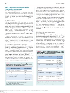
Table 16 The most commonly used simple criteria
and recognised cut-off points for definitions of electro- LVH a LV mass/BSA (g/m ) >115 (men)
2
cardiogram left ventricular hypertrophy
>95 (women)
ECG voltage criteria Criteria for LVH
LV concentric RWT >_0.43
geometry
S V1 þR V5 (Sokolow–Lyon criterion) >35 mm
LV chamber size LV end-diastolic >3.4 (men)
R wave in aVL >_11 mm
diameter/height (cm/m)
S V3 þR aVL (Cornell voltage) a >28 mm (men) >3.3 (women)
b
Cornell duration product Left atrial size Left atrial volume/height 2 >18.5 (men)
>20 mm (women)
2
(elliptical) (mL/m )
>16.5 (women)
>2440 mm.ms
BSA = body surface area; LV = left ventricular; LVH = left ventricular hypertro-
ECG = electrocardiogram; LVH = left ventricular hypertrophy.
a phy; RWT = relative wall thickness.
Sum of limb and precordial lead voltage.
a
b BSA normalization may be used in normal weight patients.
Product of Cornell voltage x QRS duration (mm.ms).
Downloaded from https://academic.oup.com/eurheartj/advance-article-abstract/doi/10.1093/eurheartj/ehy339/5079119
by guest
on 27 August 2018